
Quick links
Understanding VHL
What is VHL?
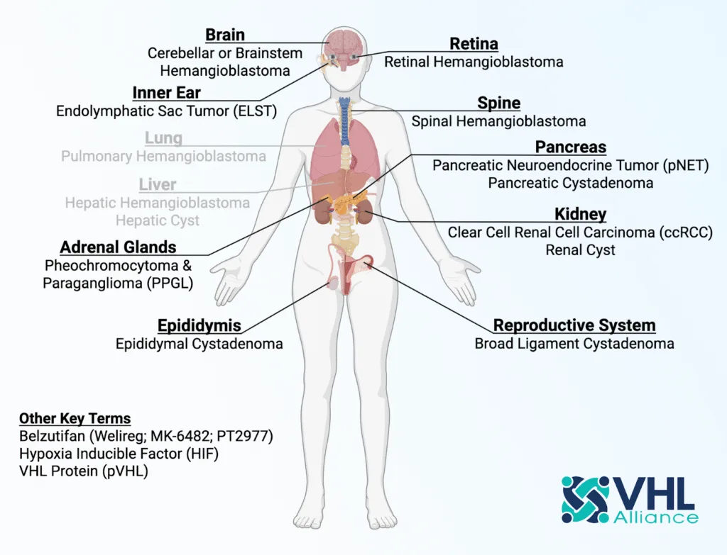
Von Hippel-Lindau Disease (VHL) is a heritable tumour predisposition syndrome caused by a flaw in a “tumour suppressor” gene– the VHL gene. “VHL” involves the growth of tumours and cysts in up to TEN organs- see the diagram above.
Tumours and cysts can form in multiple organs at the same time (multilaterally), and can also form as multiple tumours and cysts within the same organ at the same time (multifocally), and very often both multifocally AND multilaterally also at the same time
Manifestations most commonly occur commonly occur on the retina, brain and spinal cord, kidneys, pancreas, inner ears and adrenal glands
Find out more here What is VHL? Page
What causes VHL?
VHL is often inherited from a parent. About 20% of all people diagnosed with VHL have no family history of the disease (this is sometimes referred to as “de novo”), but can pass VHL on to their children.
Our bodies contain thousands of coded messages called genes that send instructions to our body on how to function; for example, what colour to make our eyes or how tall we will grow. The VHL gene makes a protein that is important for the normal function of cells. A mutation (fault) in this gene alters the message the gene sends to the body and causes an increased chance of one of the VHL tumours developing.
We each have two VHL genes, and we inherit a faulty gene from one parent, as shown in the diagram below. Statistically, therefore, we have a 50-50 chance of inheriting a faulty gene

How is VHL diagnosed?
Detection of the type of tumors specific to VHL is critical to properly diagnosing the disease. For individuals who have a family history of VHL, detection of only one type of tumor specific to VHL may be sufficient to make a diagnosis. For those patients without a family history of the disease, at least two types of VHL tumors should be identified to confirm a diagnosis. Genetic testing also may helpful in diagnosing the disease. Please contact your GP or your consultant for a blood test should you suspect you have this condition.
It is possible to look for a specific fault in the gene in an affected person. This can be done from a blood sample, since blood cells contain copies of all of our genes. This result will take a couple of months. Current techniques are not yet good enough to find all gene changes, even when the diagnosis of VHL is certain. If the fault in the gene is found, then this information can be used to test other members of the family to see whether or not they also have the faulty gene. This test is much quicker taking two to four weeks for a result.If the faulty gene has not been identified, then it may be possible to follow the gene through the family using DNA markers. This will give us a very good indication as to who is likely to carry the faulty gene within a family. Taking this information, alongside screening results, it may be possible to stop the screening programme on those people who are unlikely to have inherited the faulty gene.
Your family history may suggest there is a possibility that you have inherited VHL disease, for example children of a parent with VHL disease each have a one in two (50%) risk of developing the disease regardless of whether they are boys or girls. In the past, all children at risk were offered screening very much like that offered to people with confirmed VHL. The screening began with regular eye checks at five years and other checks were added in as the children grew into adulthood. The development of a gene test for VHL has meant that children can be tested, (usually around the age of four or five) to see whether or not they need screening. Those who test negative are not at risk of passing VHL on to their own children in the future. Those with the faulty gene will be screened appropriately. Some parents decide that they would rather the child made his or her own decision about gene testing when they are older. Therefore the child would be screened with eye checks and testing is then offered to them as they grow older and understand more about VHL and their risk. This test for children is only possible if we have been able to identify the specific VHL mutation in the family.
How common is VHL?
It is often quoted that VHL affects 1 in 36,000 people, but this figure appears to be an estimate from 1991 of “incidence” at birth, and is not a robust measure, as it largely predates genetic testing for VHL disease. A rigorous study in 2024 by Prof Eamonn Maher et al in the UK, based on 2016 population data, showed a more robust prevalence rate of circa 1 in 70,000 in the UK Learn more
Is there a cure for VHL?
Unfortunately, there is not currently a cure for VHL. Early detection and treatment of tumours significantly improves a patient’s diagnosis. Left untreated, VHL may result in blindness, permanent brain damage, permanent loss of organ function or death.
Management of VHL involves lifelong surveillance and surgeries at the appropriate times. See active Surveillance Guidelines Learn More
However, in recent years there have been major advancements in the management of VHL, belzutifan (Welireg) is approved as a “systemic” treatment in the UK for renal cell carcinoma (RCC), pancreatic neuroendocrine tumours (pNET), and CNS hemangioblastomas (HB) in adult VHL patients subject to certain criteria Learn More
Treatment and Monitoring of VHL

I have been diagnosed with VHL – who can I speak to?
This is a worrying time for anyone including carers. Sadly each diagnosis for each individual is different. Some patients experience symptoms in their teens, some don’t experience any symptoms until their 20’s or 30’s. Some don’t show symptoms at all. And even within the same family, manifestations can be completely different
We recommend you seek information and advice from your Local Genetic Centre who can provide you with information. Learn More
Follow the “Patient” tab in the Main MENU bar. for details of VHL Clinics, VHL Patient Handbook & Active Surveillance Guidelines, along with access to details of videos and events and Patient Facebook Group – Go to Patient Tab
The disease is only a part of you – it does not define you. Check out our Patient Stories page and read about other patients, carers and friends who battle with the disease every day – Go to Patient Stories
Reactions to VHL
Some people experience a range of emotions when they are told they have VHL. Anger, shock, anxiety, worry about your health, guilt about possibly passing the gene on to children are all normal reactions. Uncertainty is another problem.
Because VHL is variable, even within families, we can’t say how any one person will be affected in the future. As several family members can be affected this can also be difficult. Screening results and possible treatment interventions may be different for family members at different times. People in the family that do not inherit the gene can feel guilty when other close relatives have done so
How is VHL treated?
Complications of VHL disease are much easier to treat if they are detected early. Therefore, once a diagnosis is made, it is recommended that patients enter an annual screening programme where different parts of the body involved in VHL can be monitored See Active Surveillance Guidelines
Historically, the only available “treatment” has been surgical- to remove risky or problematic tumours.- see below. Meantime, some tumours may not be suitable for surgery, and a “systemic” drug treatment may offer a viable alternative- see Belzutifan
The following sections highlight surgical options
Hemangioblastomas of the CNS (central nervous system)
Most Brain and Spinal hemangioblastomas in patients with VHL disease can be surgically removed. Generally, they should be resected when:
- the patient becomes symptomatic; but for some patients, the window of opportunity to safely remove the tumour can be very short, or
- when the tumour has reached a size such that further growth will increase the risks associated with surgery, or
- in the event of an enlarging tumour or cyst. or
- when there is a risk to important brain tissue
Magnetic resonance imaging is usually sufficient for preoperative evaluation. and presurgical embolization is unnecessary in most (but not all) instances. The goal of surgery is complete resection of the lesion before the patient experiences a disabling neurological deficit.
Tumours can and do recur after surgery.
Gamma Knife and Stereotactic radiotherapy, offer non-invasive methods for shrinking or stabilising tumours, but do not offer a complete solution, as the tumours eventually grow again, so such treatments at best “buy time”
Hemangioblastomas of the retina / optic nerve
Hemangioblastomas of the retina are typically treated with a laser, cryotherapy, or plaque radiotherapy .
RCC (Renal cell carcinoma)
Depending on the extent of the disease, renal cell carcinoma may either be treated with
- nephron-sparing surgery (removing the tumor while sparing the unaffected portion of the kidney) or
- radical nephrectomy (removal of the whole kidney).
It is important for understand the options, and even in some cases educate your consultant on VHL , as the removal of the tumour should be done to help prevent the spread of the cancerous cells to other parts of the body, but should only be done if the tumour reaches a certain size. See Active Surveillance Guidelines
pNET (Pancreatic Neuroendocrine Tumour)
Surgery is the primary treatment modality for pNETs.
While functional pNETs should be surgically removed for symptom control as well as potential curative intent, non-functional pPNETs are increasingly managed nonoperatively. There is increasing data to suggest small, non-functional pNETs (less than 2 cm), are appropriate for nonoperative active surveillance.
- A distal pancreatectomy is used to treat pNETs present in the tail and body of the pancreas. Surgical options involve removing only the tail of the pancreas or performing a pancreatic splenectomy via open surgery or laparoscopy or robotics.
- A Whipple procedure is used to treat pNETs found in the head of the pancreas.
- A central pancreatectomy is used to treat small, low grade, tumors
Evidence supports surgical management of metastatic disease if possible, and occasionally even surgical management of the primary tumour in the setting of widespread metastases.
Pheo (Pheochromocytoma)
Minimally invasive (laparoscopic or robotic) adrenalectomy is the preferred approach for patients with pheochromocytomas. This surgical approach may be performed either through the transabdominal approach (TA) or the posterior retroperitoneoscopic approach (PRA), depending on a range of factors
VHL patients should be offered cortical-sparing adrenalectomy to maximize adrenal function, avoid chronic steroid hormone replacement, and reduce the risk of Addisonian crisis. This approach has resulted from 78% to 89% of patients with syndromic diseases and cortically-spared adrenal glands to be steroid independent at long-term follow-up
PPGL (Paraganglioma)
Pheochromocytoma and paraganglioma are together referred to as PPGL. Approximately 80%–85% of PPGL are pheochromocytomas, while about 15%–20% are paragangliomas
Surgery remains the mainstay of treatment for PPGL and offers the best chance for cure or remission. Precision surgery can be offered on an individual basis once a patient’s PPGL has been fully characterized through biochemical testing, genetic testing, anatomic imaging, and functional imaging as indicated.
Surgery recommendations from practice guidelines currently suggest laparoscopic adrenalectomy for most PHEO and open surgery for PHEO>6 cm or invasive PHEO to achieve complete resection.
Guidelines also recommend open surgery for PGL but minimally invasive techniques may be used for small non-invasive tumours in favourable locations. To date, there are no randomized controlled trials comparing Open to Minimally Invasive resection of PPGLs.
Where can I find medical professionals who are experienced with VHL?
There are a network of VHL Clinical Care Centres around the world, where patients can access coordinated care from a team of VHL experienced specialists.
To find your nearest Genetic Centre in the UK, follow the link to The VHL Clinics page HERE
What is belzutifan?
Belzutifan (Welireg) is a first-in-class targeted therapy known as a hypoxia-inducible factor 2-alpha (HIF-2α) inhibitor. It works by blocking a key protein involved in tumour growth in people with VHL. Learn more by visiting our dedicated belzutifan page HERE
What is a Hemangioblastoma?
A hemangioblastoma is a highly vascular tumour that can occur in the brain, spinal cord, and retina (the light-sensitive tissue that lines the back of the eye). Often described as “benign”, but the word “benign” is mis-leading, as a “benign” tumour that starts to grow uncontrollably within the confined space of the skull, or that becomes malignant, can be extremely dangerous and even life-threatening
While Hemangioblastomas account for about 2% of brain tumours amongst the general population, up to approximately 80% of VHL patients will develop hemangioblastomas
Within the brain, as a hemangioblastoma enlarges, it presses on the brain and can cause neurological symptoms, such as headaches, weakness, sensory loss, balance and coordination problems, vertigo and severe vomiting, and/or hydrocephalus (a build-up of spinal fluid in the brain). Some patients, however, may experience no symptoms whatsoever (they are asymptomatic), even though a tumour may be growing, But the absence of symptoms does not translate to a lack of potential harm to structures within the brain and potential loss of brain function. Hence Active Surveillance is essential
If it grows uncontrollably, the tumour may become life-threatening. These tumours are often highly vascular (connected to lots of blood vessels), and therefore surgery itself could be life-threatening.
Retinal hemangioblastomas (R-Hb’s) are always benign. Circa 90% of VHL patients will experience R-Hb’s. Their impact is the risk of blindness due to detachment of the retina, or if they are located at the optic disc where treatment options like proton beam radiation are limited and always associated with loss of vision
Spinal cord hemangioblastomas in VHL can be complex, due to the frequent multiplicity and variable nature of these tumours along the craniospinal axis. Nevertheless, surgical resection is the treatment of choice, where viable
What is ccRCC?
ccRCC is known as clear cell Renal Cell Carcinoma and is the most common type of kidney cancer. It affects the tubules that filter waste from your blood. The cancer cells look like clear bubbles under a microscope. Clear cell renal cell carcinoma cells rapidly multiply, forming one or more tumours. It usually only affects one kidney, but can affect both.
Generally, VHL patients have a 70% chance of developing ccRCC
What is a pNET?
Around half of people with VHL may have cysts in the pancreas, which don’t usually cause any symptoms. However, some cysts can become cancerous, so cysts should be assessed to check the risk of them becoming cancer (pNET)
The prevalence of pancreatic neuroendocrine tumours (PNETs) in VHL disease ranges from 9% to 17% and is characterized by a better prognosis compared with sporadic pNET’s. These tumours are usually non-functioning. This means they don’t produce more hormones than normal and generally don’t cause hormone-related symptoms.
Several risk factors for PNET metastasis in VHL disease have been reported but have not been validated
Even if not classified as pNET’s some cysts can block the pancreas and cause a range of serious problems, all requiring surgery
What is a pheo?
A phaeochromocytoma (“pheo”) is a rare tumour of the adrenal glands, which sit above the kidneys. Generally, VHL patients have a 20% chance of developing a pheo; However this can be a high as 70% for VHL Type 2 patients
Most pheos do not spread to other areas of the body, but around 1 in 10 do. These are called metastatic phaeochromocytomas.
It’s usually possible to successfully remove a phaeochromocytoma using surgery.
What is a PPGL?
A paraganglioma (also known as an extra-adrenal pheochromocytoma) is a rare neuroendocrine tumour (NET) that forms near your carotid artery (the major blood vessels in your neck), along nerve pathways in your head and neck and in other parts of your body. The tumour is made of a certain type of cell called chromaffin cells, which produce and release certain hormones known as catecholamines.
Even though paragangliomas don’t form in your adrenal glands, they’re made of tissue that’s found in your adrenal glands
Your adrenal glands, the two glands that are on top of each kidney, make several hormones. Among these are hormones called catecholamines that help control the following important bodily functions:
• Heart rate.
• Blood pressure.
• Blood sugar (blood glucose).
• The way your body responds to stress.
Clinicians

I am a Medical Professional – How can I connect my patient with the charity?
VHL UK/Ireland is run by volunteers and we have setup a fantastic network of support within the charity for patents and carers.
To get in touch just click here Contact Us
Don’t forget our social media pages too- Links are on the foot of any page you visit within the VHL UK/Ireland website.
We also have a Facebook Group where there is a fantastic support network of #vhlwarriors – Go to Facebook Group
I am a Medical Professional – how can I be made aware of future VHL events to communicate to patients?
Please join our mailing list so that we may send notifications of upcoming events JOIN HERE
Please also check out our Events Page HERE
Don’t forget you can Contact-Us for any specific questions.
Research
What is being done about research?
Find out by following this link to our Research page HERE
About the Charity

What is the VHL Alliance?
The VHL Alliance (VHLA) is a 501c3 non-profit organization founded in 1993 by three families with VHL to share experiences, learn from and support from one another, and help doctors understand and treat VHL to improve life for patients. The VHL Alliance provides significant resources for patients, caregivers, researchers, and the medical community.. Whilst its primary focus is to support VHL patients in North America, it also helps patients in other countries to establish their local country charities / patient organisations
What is the VHL UK/Ireland Charity?
VHL UK/Ireland Charity supports VHL patients in the UK & Ireland, and also supports patients with two other genetic conditions (specifically BHD and HLRCC) which exhibit SOME manifestations which are similar to those experienced by VHL patients. It does this, working in collaboration with the VHL Alliance, by:
- Providing support and information for patients regarding their genetic conditions
- Providing funds for research into the genetic conditions and for equipment to assist with such research
- Advancing the education of the public in all matters concerning these genetic conditions
How can I get involved with the Charity?
There are lots of ways you can get involved- Check out our Volunteer Page HERE
Supporting Us

How can I fundraise for VHL?
There are many ways to help raise awareness and fund-raise for VHL UK/Ireland. Please visit our fundraising page for the most up to date information on fundraising, where you can create your sponsorship page and fundraising downloads such as sponsorship forms etc.
I have completed my fundraiser – how do I send you the money raised?
If you have used an online sponsorship page, funds will automatically be paid to the charity.
However you may receive cash / bank transfer donations and cheque donations.- you can get in touch for bank details using the Contact US page-Just click Contact-Us
Let us know if you need the UK or Ireland banking details to transfer to. We advise to use a bank transfer as we do not incur any costs, but if it is easier you can PayPal them across.
Cheques: payable to VHL UK/Ireland can be posted to VHL UK/Ireland, 47 Tiln Lane, Retford, Notts, DN22 6SN.
How can I get involved with the charity?
There are lots of ways you can get involved- Check out our Volunteer Page HERE